Digital surgical guides have emerged as a transformative “high-precision navigator” in modern implant dentistry, revolutionizing how dental implants are placed by minimizing surgical errors to the millimeter scale. As a patient-specific auxiliary device, this technology bridges virtual treatment planning and clinical execution, ensuring that implant position, angle, and depth align perfectly with both anatomical constraints and prosthetic requirements.
The core workflow of digital guided implant surgery embodies the integration of advanced imaging and additive manufacturing. It begins with high-resolution data acquisition: Cone Beam Computed Tomography (CBCT) captures 0.1mm-level 3D volumetric data of the jawbone, clearly visualizing tiny neurovascular bundles and sinus cavities that were once challenging to assess with 2D radiography. AI-powered software (such as Codiagnostix™ and Implant Studio) then fuses these CBCT data with intraoral scan results, allowing clinicians to simulate the entire implant process virtually—adjusting implant specifications based on bone density and avoiding critical anatomical structures. The finalized plan is translated into a physical guide via high-precision stereolithography 3D printing, using medical-grade resins with flexural strength up to 120MPa to withstand surgical drilling forces.
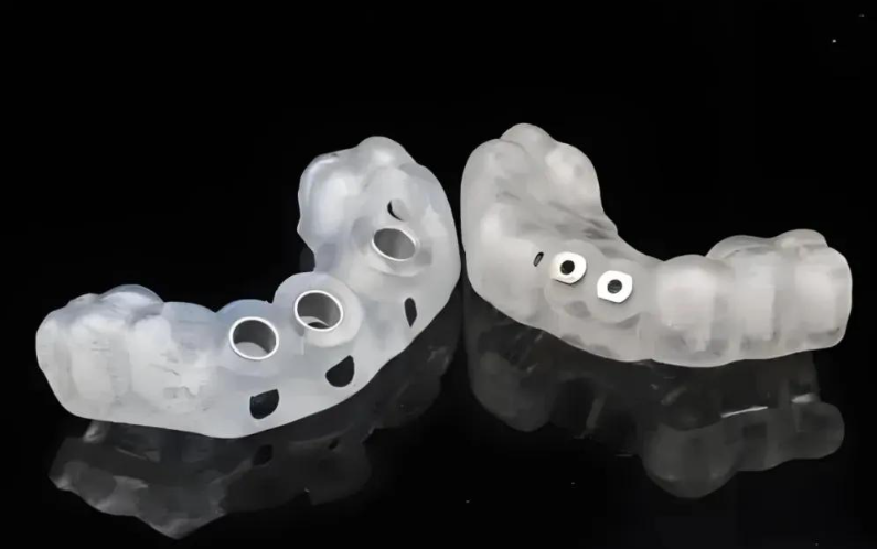
These guides are categorized by their support mechanisms to adapt to diverse clinical scenarios: tooth-supported guides for patients with stable adjacent teeth, bone-supported variants for edentulous areas requiring direct skeletal fixation, mucosa-supported designs for preliminary assessments, and hybrid models combining multiple stabilization methods. Regardless of type, the embedded metal guiding sleeves act as “fixed rails” during surgery, ensuring drills and implants strictly follow the pre-planned path—reducing average coronal deviation to 0.73±0.53mm and apical deviation to 1.16±0.62mm.
The clinical advantages of this technology are multifaceted. Safety is significantly enhanced: by avoiding inferior alveolar nerves and maxillary sinuses, complications like nerve injury are drastically reduced, and 1-year implant survival rates reach 98.98%—with some long-term studies reporting 100% retention over a decade. Minimally invasive procedures become standard: 2-3mm incisions replace traditional 1-2cm flaps, cutting single-implant surgery time from 40 minutes to 15 minutes and full-arch treatment from 8 hours to 2.5 hours. Patient outcomes are also improved aesthetically and functionally: precise implant positioning optimizes prosthetic load distribution, achieving a pink and white esthetic score of 9.53±1.68 and overall patient satisfaction of 9.15±0.84.
Recent advancements continue to expand the technology’s potential. Dynamic navigation systems like X-Guide® integrate real-time optical tracking with digital guides, compensating for intraoperative patient movement and providing turn-by-turn drill guidance without relying on physical templates. Machine learning algorithms now predict treatment outcomes by analyzing 38 clinical variables, with a prediction accuracy of 91.7%—particularly valuable for high-risk cases like smokers or patients with type II bone density.
In essence, digital surgical guides transform implant dentistry from an experience-dependent practice to an evidence-based discipline. By translating complex anatomical data into actionable precision, they empower clinicians to tackle challenging cases confidently while delivering faster recovery, fewer complications, and more predictable results—redefining the gold standard for modern oral rehabilitation.